Plain X ray abdomen showing a dilated stomach with nasogastric tube in situ

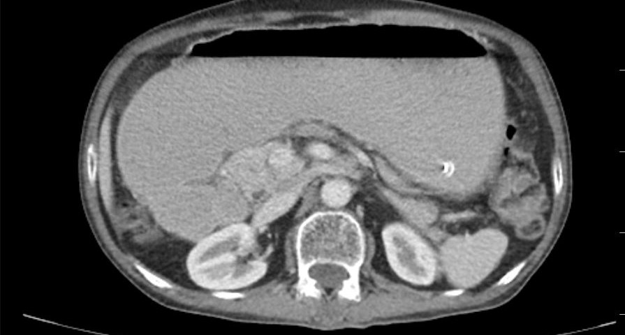
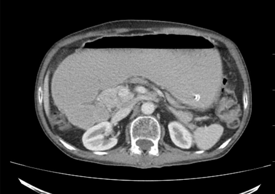
CT scan abdomen showing dilated stomach and proximal duodenum

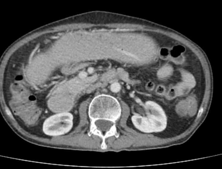
CT scan showing obstruction at level of third part of duodenum without any obvious mass lesion

An Unusual Case of Duodenal Obstruction
A 61 year old male presented to us with history of epigastric fullness after meals of 6months duration. His symptoms progressively worsened and he had recurrent episodes of bilious vomitingsince 15 days prior to of presentation. At the time of review in our OPD he was unable to tolerate any solid food. He was dehydrated and his stomach was grossly dilated. CECT abdomen was done which revealed afocal narrowing in the third part of the duodenum with significant dilation of the proximal duodenum and stomach. There was no definite mass lesion but minimal surrounding fat stranding seen along withfew enlarged lymph node. UGI endoscopyshowed a lot of fluid in the stomachwith patulous pylorus and narrowing the third part of duodenumwhich appeared like a diaphragm. Considering his age and clinical presentation, a malignant pathology was suspected and he planned for surgery after optimization.
- On exploration, there was astricture in the middle of the third part of duodenum with proximal dilation. Proximal duodenum was thickened and inflamed while distal duodenum and jejunum were normal. A segmental resection of the affected part with a Roux-en-Y gastrojejunostomywas done. Biopsy report revealed the lesion to be ulceroinflammatoryin nature with no evidence of malignancy. Postoperatively the patient recovered well and was discharged 6days after surgery by when he was tolerating normal diet.


