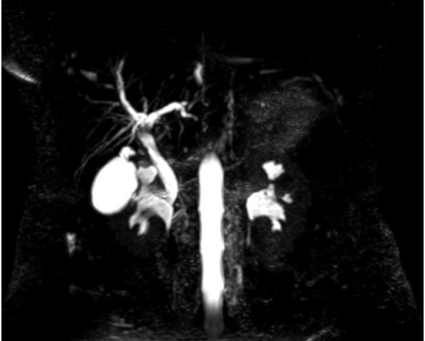
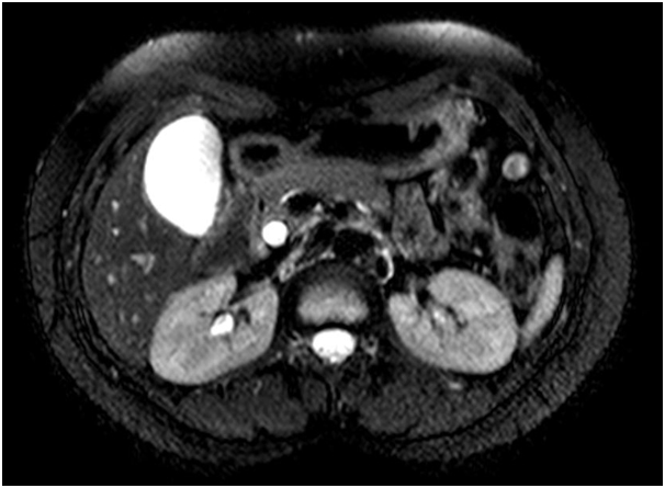
MRCP-showing-suspicious-filling-defects-in-CBD
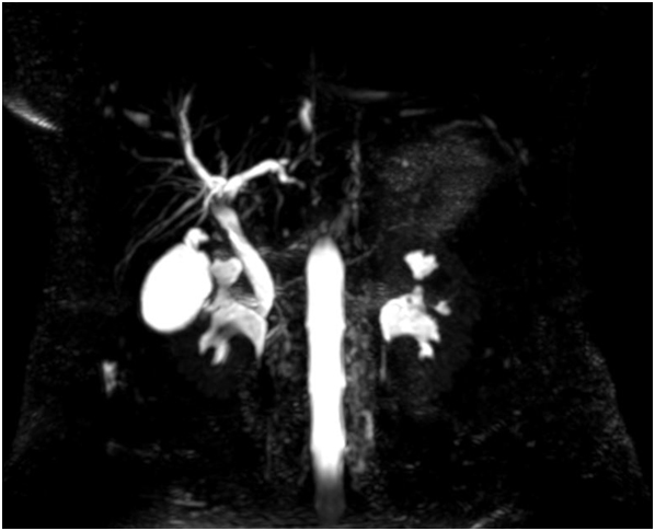
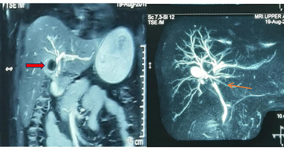
MRI-showing-dilated-CBD
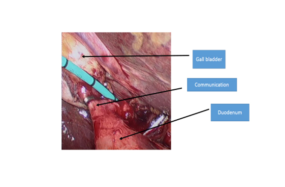
Operative-Photograph-showing-communication-between-gall-bladder-and-duodenum-with-catheter-in-cystic-duct-(for-cholangiogram)
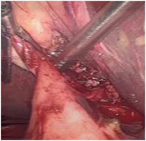
Operative-photograph-showing-fistula-being-divided
Operative-Photograph-with-clip-placed-across-the-fistula
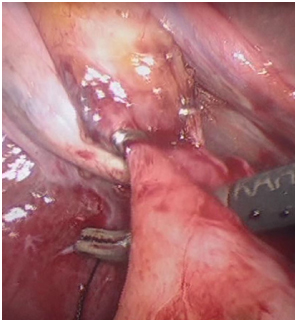
A 39 years old female was admitted with complaints of recurrent pain abdomen of many years duration. She gave a history of jaundice in the past (1998). She also gave a history of fever with chills one month prior to presentation to the OPD. Her USG abdomen revealed presence of stones in the gall blabber and with a dilated CBD about 10mm in size. MRI with MRCP was done which confirmed stones in the gall bladder and also revealed small filling defects in the CBD suggestive of small calculi. An ERCP done by the medical gastroenterologist revealed sludge in the CBD. Papilotomy was done and CBD clearance done with a balloon catheter. She was then planned for laparoscopic cholecystectomy. At laparoscopy the anatomy raised suspicion of a cholecystoduodenal fistula (communication between gall bladder and small intestine). A laparoscopic cholangiogram was done to further clarify the anatomy. The fistula was dealt with by division of the fistula and repair of duodenum by laparoscopic suturing. Postoperatively patient did and was discharged 2 days after the procedure.
Public interest: cholecystodudenal fistula is a rare complication of cholelithiasis which can be dealt with laproscopy in the hands of advanced laparoscopic surgeons.


